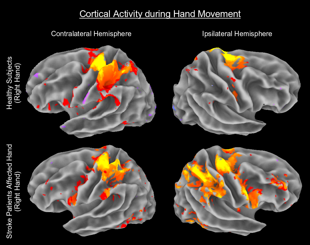
Left to right) Sagittal T2-weighted brain magnetic resonance imaging... | Download Scientific Diagram

Patient 2's magnetic resonance imaging scan; left and right refer to... | Download Scientific Diagram
An Evaluation of the Left-Brain vs. Right-Brain Hypothesis with Resting State Functional Connectivity Magnetic Resonance Imaging | PLOS ONE
![PDF] Normal human left and right ventricular and left atrial dimensions using steady state free precession magnetic resonance imaging. | Semantic Scholar PDF] Normal human left and right ventricular and left atrial dimensions using steady state free precession magnetic resonance imaging. | Semantic Scholar](https://d3i71xaburhd42.cloudfront.net/a115a025f7655b26dbcf8d60f6892370f84ef679/3-Figure1-1.png)
PDF] Normal human left and right ventricular and left atrial dimensions using steady state free precession magnetic resonance imaging. | Semantic Scholar

Axial (left, center) and coronal (right) T1-weighted magnetic resonance... | Download Scientific Diagram
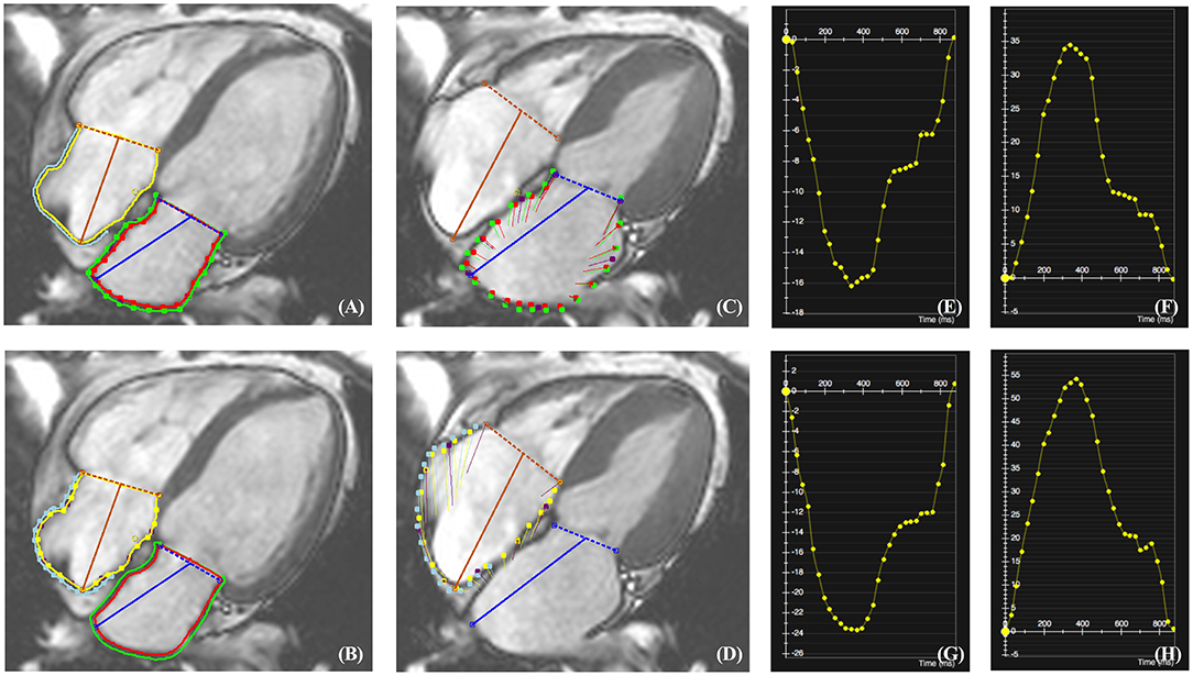
Frontiers | Quantitative Assessment of Left and Right Atrial Strains Using Cardiovascular Magnetic Resonance Based Tissue Tracking

t1-(right) and t2-(left) weighted magnetic resonance imaging images... | Download Scientific Diagram

Patient 2's magnetic resonance imaging scan; left and right refer to... | Download Scientific Diagram
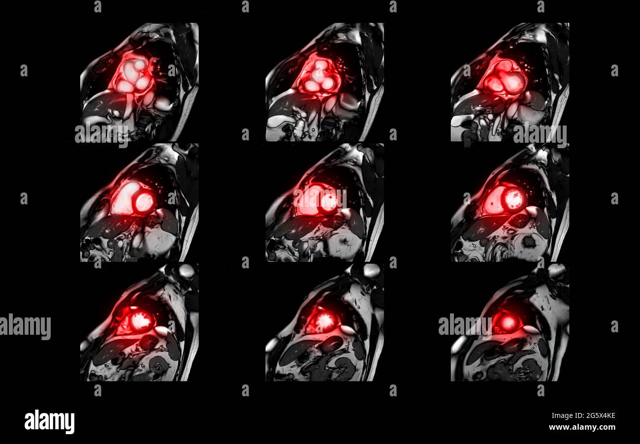
MRI heart or Cardiac MRI magnetic resonance imaging of heart in short axis view showing cross-sections of the left and right ventricle for detecting h Stock Photo - Alamy
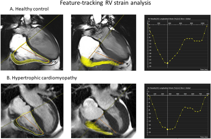
Right ventricular function declines prior to left ventricular ejection fraction in hypertrophic cardiomyopathy | Journal of Cardiovascular Magnetic Resonance | Full Text

Left, right, or bilateral amygdala activation? How effects of smoothing and motion correction on ultra-high field, high-resolution functional magnetic resonance imaging (fMRI) data alter inferences - ScienceDirect
60773-5.fp.png)
PROGNOSTIC VALUE OF RIGHT VENTRICULAR EJECTION FRACTION DETERMINED BY CARDIAC MAGNETIC RESONANCE | Journal of the American College of Cardiology

The fundamentals of left ventricular assessment in cardiac magnetic resonance imaging (CMR) - YouTube
![PDF] Human taste cortical areas studied with functional magnetic resonance imaging: evidence of functional lateralization related to handedness | Semantic Scholar PDF] Human taste cortical areas studied with functional magnetic resonance imaging: evidence of functional lateralization related to handedness | Semantic Scholar](https://d3i71xaburhd42.cloudfront.net/831eace0dc8934345f9d79917b953e689056a65d/2-Figure1-1.png)
PDF] Human taste cortical areas studied with functional magnetic resonance imaging: evidence of functional lateralization related to handedness | Semantic Scholar
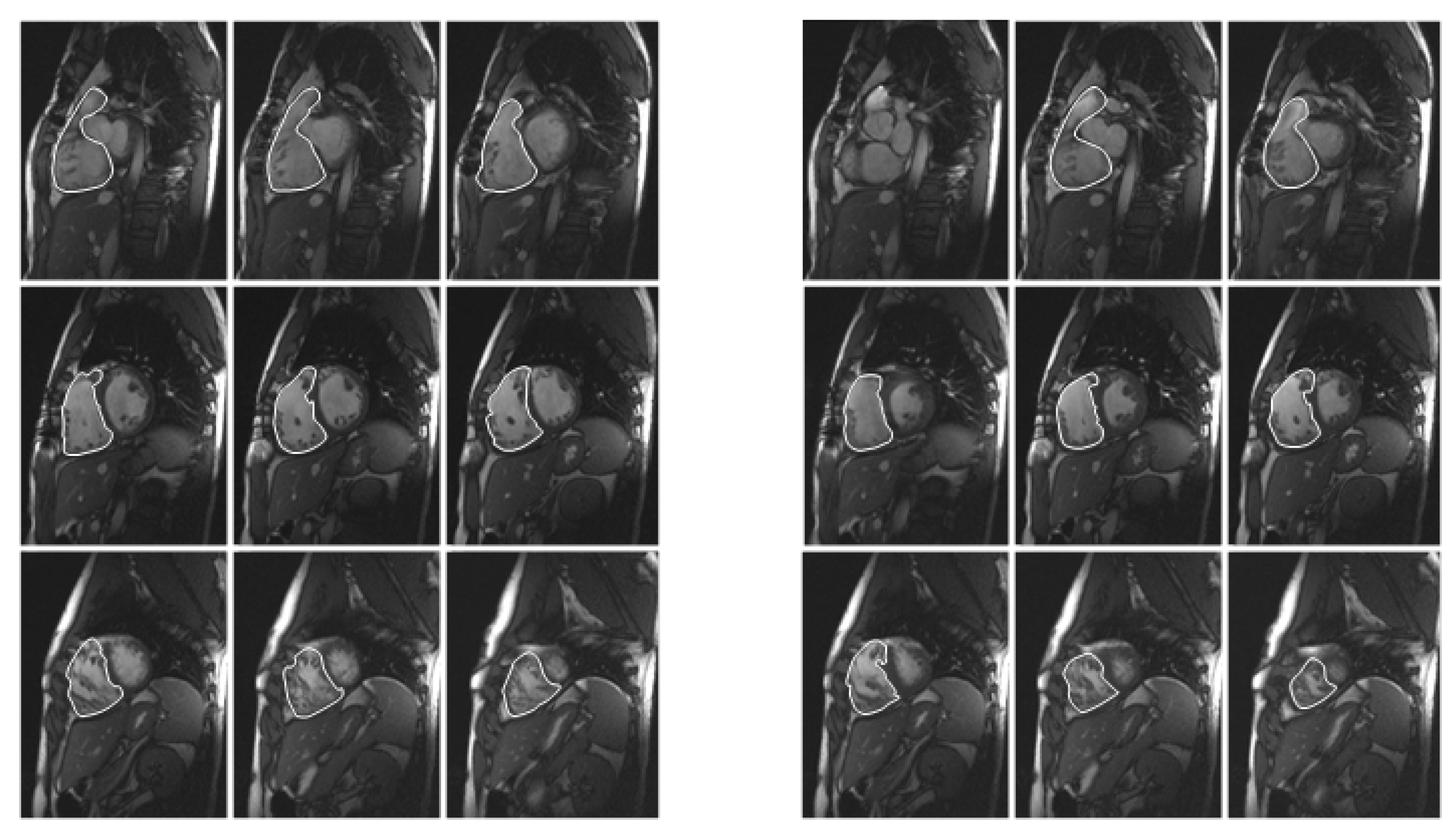
Diagnostics | Free Full-Text | Quantification of Right and Left Ventricular Function in Cardiac MR Imaging: Comparison of Semiautomatic and Manual Segmentation Algorithms

The Spatial Distribution of Late Gadolinium Enhancement of Left Atrial Magnetic Resonance Imaging in Patients With Atrial Fibrillation | JACC: Clinical Electrophysiology

A preferred patient decubitus positioning for magnetic resonance image guided online adaptive radiation therapy of pancreatic cancer - Physics and Imaging in Radiation Oncology
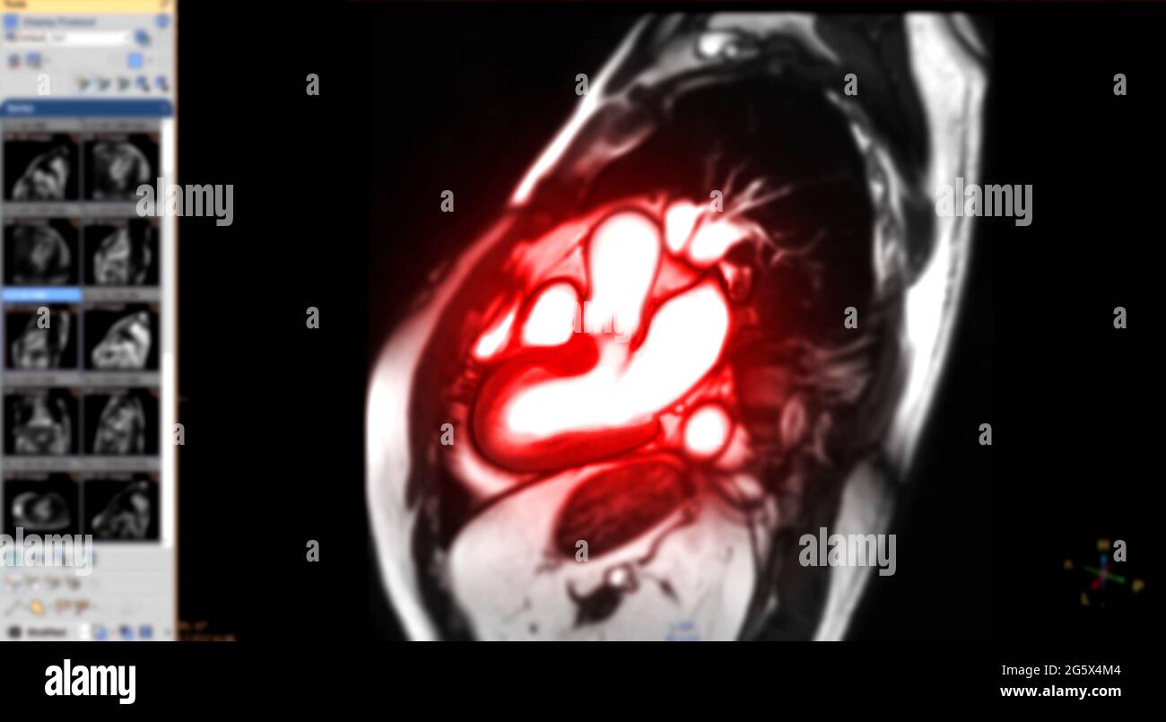
MRI heart or Cardiac MRI magnetic resonance imaging of heart in Sagittal view showing cross-sections of the left and right ventricle for detecting hea Stock Photo - Alamy

